IVF pregnancy ultrasound at 5 weeks 2 days
Five week pregnancy ultrasound with sac and yolk sac
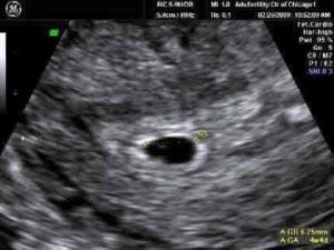
Transvaginal ultrasound, normal pregnancy at 5 weeks 2 days
Gestational sac (black area ) and yolk sac are seen
Sac measures 6.25mm diameter
Yolk sac (small white circle in left side of the sac)
Yolk sac is a source of nutrients for the fetus
The fetus is too small to be seen this early in pregnancy
Identical twin pregnancy ultrasound
Ultrasound of another pregnancy at same gestational age as above
This shows identical (monozygotic) twins – 1 gestational sac with 2 yolk sacs visible inside it
This results from an early embryo dividing into 2 genetically identical embryos
The vast majority of twins from assisted reproductive technology procedures are not “identical”, but result from implantation of 2 embryos that were the result of fertilization of 2 different eggs, as shown in the ultrasound below.
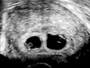
Ultrasound of a twin pregnancy
Fraternal twins result from implantation of 2 genetically different embryos
Yolk sacs and fetal poles (the early fetus) are seen in 2 completely separate sacs
This pregnancy is at 6 weeks