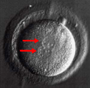
Picture of IVF embryo – this is a fertilized human egg (also called oocyte). This is the morning after IVF egg retrieval when we check eggs for fertilization. Male and female genetic material (DNA) are in the 2 pronuclei –(red arrows).
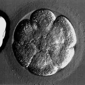
Photo of a high quality day 3 human embryo at the 8-cell stage 6 cells are visible in this plane of focus
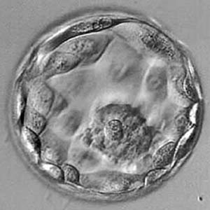
This picture shows a high quality blastocyst embryo on day 5 5 day blastocyst transfer can result in high pregnancy rates with low multiple pregnancy rates
Pictures of eggs and sperm Human eggs, also called oocytes

Eggs as seen at the egg retrieval procedure The eggs (darker area below the “E”) are inside a mass of cumulus cells (“C”)
Images showing examples of normal and abnormal eggs, including low quality eggs, high quality mature eggs, abnormal eggs, very immature eggs, and degenerative oocytes.
Human sperm pictures
In the end, us men are just DNA donors. Some women would (? falsely) claim that’s all we’re good for! If cloning is ever perfected and applied to humans, women won’t even need us for our DNA. Let’s hold off on the cloning research, please…
Pictures of day one embryos
Fertilized human eggs, one-cell embryos, also called 2 pronuclear embryos or zygotes
This is what we want to see the morning after the egg retrieval in IVF cycles
Embryos such as this must be discarded.
Pictures of day 2 embryos
This is what we want to see at about 48 hours after the egg retrieval procedure
Two embryos are shown, one is multinucleated and very abnormal, the other appears normal 2-cell embryo photos: Fragmentation
Two embryos are shown, one has some fragmentation, the other does not
Images of day three embryos
This is what we want to see at about 72 hours after egg retrieval Embryos that look like this generally have a high rate of implantation after being transferred to the uterus
Pictures of normal and abnormal day 3 embryos
One embryo has a very unusual shape. Is it to become a football star? An example of a very fragmented and low quality embryo is also shown.
Pictures of day four morula stage embryos
Embryo images at the morula stage
This is the last stage of embryo development before it (hopefully) becomes a blastocyst
Pictures of blastocysts on day 5 to 6 of IVF embryo development
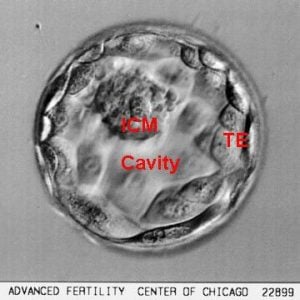
The blastocyst stage is the last stage of embryo development before the embryo hatches and implants into the lining of the uterus. Blastocyst transfer can reduce multiple pregnancy risks, with high pregnancy success rates.