Uterine polyps sometimes cause problems with fertility – or increase the risk of miscarriage.
A “normal” uterine cavity and endometrial lining is needed to conceive and maintain pregnancy.
What is a uterine polyp?
A polyp is an overgrowth of tissue in the lining (endometrium) of the uterus. The concept is similar to that of a skin tag – basically normal tissue, but growing in an abnormal formation.
Many polyps are very small (a few millimeters in diameter) and do not represent a compromise to reproductive capabilities. However, large polyps – or multiple polyps – can interfere with reproduction by causing infertility, or by increasing risks for miscarriage.
Ultrasound pictures with polyps:
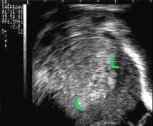
Picture at left is ultrasound of a uterus with polyps in the endometrial lining (between green L’s). The polyps can not be seen well because they are hidden in normal endometrial tissue.
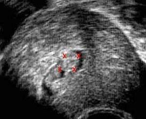
Photo at right shows the same uterus with sterile salt water injected in the uterine cavity. This test is called a sonohysterogram, or hydrosonography. The polyps are surrounded by water (black) and are easily seen – one is between the red X’s.
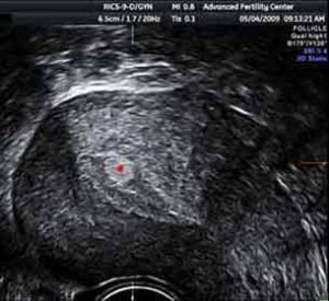
Ultrasound picture shows uterine lining with polyp (red dot at middle of polyp)
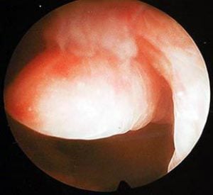
Hysteroscopy image of the polyp at surgery to remove it
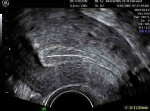
Normal uterus on ultrasound
For comparison, an ultrasound picture of a uterus with a normal endometrial lining
Cursors at top and bottom of lining show that it is 11.2mm thick
Hysteroscopy Pictures Showing Uterine Polyps
Hysteroscopy is done by inserting a narrow scope through the cervical opening allowing us to see in the uterine cavity. Instruments can then cut and remove abnormal tissue.
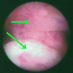
2 polyps (green arrows) discovered in uterine cavity with office hysteroscopy
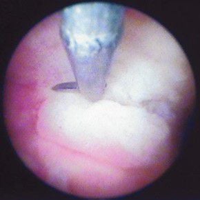
Polyp being resected with scissor and office hysteroscopy
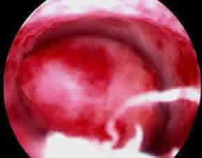
A large polyp took up most of uterine cavity
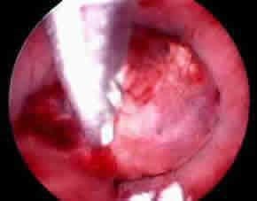
Hysteroscopic scissors resecting the polyp